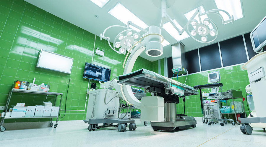
In vivo testing in patients could contribute to taking a new step forward towards increasingly sophisticated and optimised “precision surgery” on the patient. The testing was launched in recent weeks to validate a radioguided surgery technique with drugs that emit beta radiation. The new technique, developed by the Sapienza University of Rome and INFN, is the fruit of the close interdisciplinary collaboration between physicists, chemists, radio-pharmacists, nuclear physicians and surgeons, and could become an additional tool to support the oncological surgeon when removing tumours.
Radioguided surgery is a technique that makes it possible to identify residual tumours in real time. The technique consists in detecting, thanks to a probe, the radiation emitted by a radioactive substance, a radiopharmaceutical containing a specific molecule that is recognised and metabolised by the receptors of tumour cells. In this way, it is possible to directly verify, during surgery, whether the tissue analysed is a tumour or not, and, thus, guide the surgeon on the sites to be removed. The project is based on an initial idea, patented in 2013 by Sapienza, INFN, and the Enrico Fermi Research Center, which involved the use of beta- radiation, a type of radiation that poses problems in application due to the limited availability of radiopharmaceuticals with this type of emission. Thus, after studies conducted in collaboration with the “Carlo Besta” Neurological Institute, the European Institute of Oncology, the Leiden University Medical Center, and the Agostino Gemelli University Hospital Foundation, the choice fell on beta+ radiation, used on a daily basis in nuclear medicine departments for PET (Positron Emission Tomography) diagnostic testing. In any case, despite their wide availability compared to those emitting beta, these radiopharmaceuticals have difficulties linked to the abundance of photons produced not just in the diseased tissues, but also in all the areas of the body reached by the molecule after administration, which may disturb the signals detected by the probe. For this reason, it is necessary to continue with tests that make it possible to understand and calibrate the device and provide doctors with indications, for example, on the levels of counts associated with the actual presence of a tumour. After years of feasibility studies and ex-vivo tests, i.e. on tissue samples removed from patients undergoing surgery, recent testing is now underway, with the prototype developed by NUCLEOMED Srl, at the European Institute of Oncology in Milan, where the potentials of this technique in both neuroendocrine tumours of the gastrointestinal tract and prostate carcinomas are studied in detail, and at the Molinette Hospital of “Città della Salute di Torino”, concerning prostate tumours.